Summary
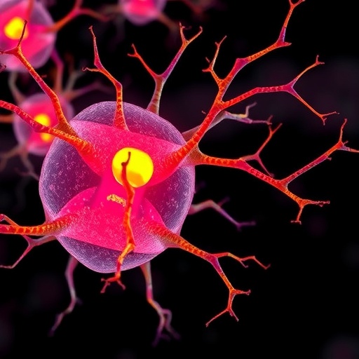
In an era where metabolic diseases are rapidly becoming a pressing global health concern, researchers are unveiling the intricate biological mechanisms that underpin these conditions. A recent study by Ercin and Gezginci-Oktayoglu sheds light on the pivotal role of cellular plasticity in metabolic d…
Source: BIOENGINEER.ORG

AI News Q&A (Free Content)
Q1: What is the role of cellular plasticity in metabolic dysfunction-associated steatosis as suggested by recent research?
A1: Recent research indicates that cellular plasticity plays a significant role in metabolic dysfunction-associated steatosis. Hepatocytes, under chronic high insulin stimulation, may dedifferentiate into liver progenitor cell-like populations. This transformation is further influenced by exposure to fatty acids, leading to the formation of adipocyte and fibroblast-like cells. This cellular transformation contributes to lipid accumulation and the progression of metabolic steatosis.
Q2: How does chronic high insulin exposure affect hepatocytes according to the latest studies?
A2: Chronic high insulin exposure causes hepatocytes to dedifferentiate, forming liver progenitor cell-like populations. This process is marked by increased expression of progenitor cell markers and reduced expression of typical hepatocyte markers. Such dedifferentiation contributes to the development of adipocytes and fibroblast-like cells, facilitating lipid accumulation and metabolic disturbances.
Q3: What molecular mechanisms are involved in the dedifferentiation of hepatocytes under prolonged insulin and fatty acid exposure?
A3: The dedifferentiation of hepatocytes under prolonged insulin and fatty acid exposure involves several molecular mechanisms, including the upregulation of CD34 and PDGFR1α markers. Inhibitors targeting TLR4 and GSK3β have been shown to reduce lipid accumulation, while the suppression of TLR4 and β-catenin helped prevent the increase in fibroblast-like cells. This indicates a complex interaction between signaling pathways in driving cellular plasticity and steatosis.
Q4: What are the implications of lipid accumulation in cells due to cellular plasticity for metabolic diseases?
A4: Lipid accumulation in cells, driven by cellular plasticity, is a critical factor in the development of metabolic diseases such as steatosis. The transformation of hepatocytes into progenitor-like cells and subsequent adipogenesis exacerbate lipid storage in liver cells, contributing to metabolic imbalances and disease progression.
Q5: How does cellular plasticity contribute to the development of adipocyte and fibroblast-like cells in liver tissues?
A5: Cellular plasticity contributes to the development of adipocyte and fibroblast-like cells through the dedifferentiation of hepatocytes, which is stimulated by prolonged high insulin and fatty acid exposure. This process increases the expression of progenitor and fibroblast markers, enabling the transformation of liver cells into adipocyte-like and fibroblast-like phenotypes.
Q6: What role do TLR4 and GSK3β inhibitors play in addressing lipid accumulation in liver cells?
A6: TLR4 and GSK3β inhibitors play a crucial role in mitigating lipid accumulation in liver cells. By targeting these pathways, the inhibitors help reduce the dedifferentiation and transformation of hepatocytes into lipid-storing adipocyte-like cells, thus addressing one of the key pathological features of metabolic steatosis.
Q7: How does exposure to oleic acid following insulin impact fibroblast marker expression in liver cells?
A7: Exposure to oleic acid following insulin treatment increases the expression of fibroblast markers in liver cells. This is indicative of a shift towards fibroblast-like phenotypes, contributing to the fibrotic changes observed in metabolic steatosis. The increase in fibroblast markers suggests a complex interplay between lipid exposure and cellular plasticity in disease progression.
References:
- Exploring the role of cellular plasticity in metabolic dysfunction-associated steatosis and related molecular mechanisms
- Cell (biology)
- Atherosclerosis


